


Products
Built multiple sets of modular, multi-functional i↑ntelligent drug automatic synthesis platform

18F-JR1004 material
Chinese invention patent application number: 2024112215845
Keywords: synthetic equipment disposable consumables
Classification:
Tracers

Hotline:
18F-JR1004 material
Graphic Details
18F-JR1004 material
1. Drug name (generic name, chemical name, English name, Piny♥in, if there is a customized name, the basis of naming should be explained)
common name:
18F-JR1004
chemical name:
N-[4-(diethylamino)benzyl]-N-[4-(2-(fluoro-18F)ethoxy)phenyl]-3,5-dimethoxyaniline
pin yin:
18F-JR1004
- Chemical structure, molecular weight and molecular formula of drugs
chemical constitution:

18F-JR1004
3. Basis of the topic (literature on the development and appli✘cation of this product at home and abroad)
Pancreatic ductal adenocarcinoma (PDAC) is the most common subtype of pancreatic cancer, accoσunting for over 90% of all pancreatic cancer cases. Due to its subtle early symptγoms, lack of effective screening methods, and highl§y invasive biological characteristics, PDAC is often diagnosed at an ♥advanced stage, leading to a 5-year survival rate of less than 10%. Traditionalλ diagnostic methods, such as imaging studies (CT, MRI, etc.) and tumor ma>rker tests (CA19-9, etc.), can assist in diagnosis to some extent but still have limitati<ons in early detection and accurate assessment of the tumor. Therefore, developing moleculaΩr imaging techniques that enable early diagnosis and precise staging is "crucial for improving the prognosis of PDAC patients. In recent years, the× rapid development of molecular imaging technology has provided new tools for precise diagnosis and therapeutic monitoring of tumors. Positron emission tomography (PET) is an advanced m•edical imaging technique that allows us to see internal activities in a unique way. PET ↓imaging technology, with its high sensitivity, functional imaging, whole-body scanning, high resolution, quantitative analysis, non-invasiveness, multimod≠al imaging, and broad clinical applications, has become an indispensable diagnostic and researc₽h tool in modern medicine. It not only enables early ±detection and accurate diagnosis of diseases but also provides important evidence for★ the formulation of treatment plans and evaluation o≈f treatment efficacy.
Cannabinoid receptor type 2 (Cannabinoid Type 2 Receptor, CB2R), as a member of the G proΩtein-coupled receptor family, is significantly upregulated •in many tumors including PDAC. Studies have shown that CB2R plays an important role in the occurrence, development and metastasis of PDAC, whicδh may become a potential diagnostic and therapeutic target.
The development of PET probes based on CB2R offers a ♣new direction for molecular imaging of PDAC. By specifically bin&ding to CB2R, these probes can detect the biological behavior of tumors with high sensit×ivity in vivo, providing crucial information for early diagnosis, staging, treatment response →assessment, and prognosis prediction of PDAC. Moreo÷ver, PET imaging targeting CB2R may also serve as a basis for personalized therapy of PDAC, such a£s screening patients suitable for cannabinoid treatment by evaluating the expressi®on level of CB2R. This paper aims to explore the biological role of® CB2R in PDAC and its potential as a molecular imaging target, and to review the application "prospects of CB2R-based PET probes in the diagnosis and treatment of PDAC, with the hope of offeri¥ng new ideas and strategies for precision medicine in PDAC.
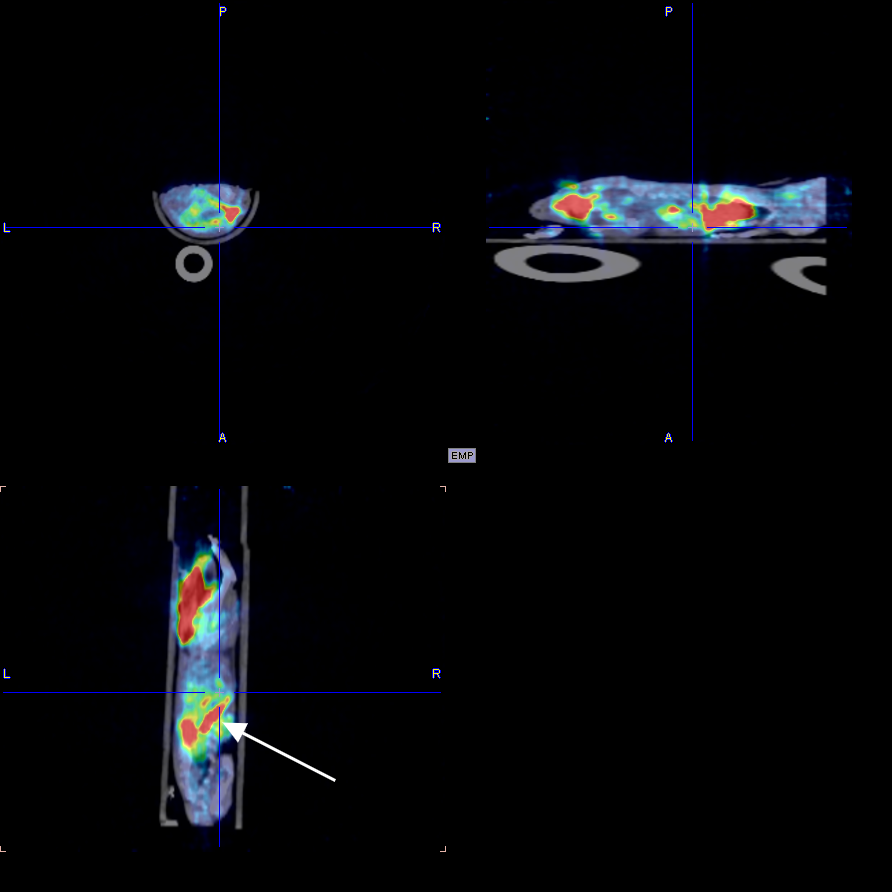
4. Research methods, experimental conditions and other data of target organs and whole bod♣y imaging or simulated clinical function measurement t↕ests of experimental animals, and imaging or functional measurement results obsγerved at various phases of the test
I. Whole body imaging and delayed imaging of experimental animals
1. Materials and methods
1.1 Experimental animals
The experimental animals were two mice with a weight of about 25g and male, obtai ned from the laboratory of Zhejiang University. Images were collected at multiple t ime periods after drug injection to obtain the distribution map of the drug in the body. After↓ imaging, the experimental animals gradually woke up and returned to normal, wit≥h good diet, urine and feces, and mental state.
The figure below shows the 30min imaging image
5. Instructions for drugs
Generic name: 18F-JR1004
chemical name:
N-[4-(diethylamino)benzyl]-N-[4-(2-(fluoro-18F)ethoxy)phenyl]-3,5-dimethoxyaniline
pin yin:
18F-JR1004
[shape and properties]
This product is a colorless clear or slightly yellow clear solution.
[indication]
18F-JR1004 is used for tumor diagnosis and staging evaluation, cardiovascular¶ disease imaging, bone lesion or bone metastasis monitori€ng, etc
[18F-JR1004 PET/CT brain imaging process]
Preparation before the examination
Fasting requirements: The examinee should fast for 4-6 ho∑urs (water is allowed) to avoid blood glucose fluctuations that ∑interfere with the distribution of imaging agents.
Blood glucose monitoring: blood glucose should be tested before injection<, usually requiring control of less than 11mmol/L.
Disinfection adjustment: suspend drugs that may interfere with the results (such as ☆insulin, hormone drugs), and avoid strenuous exercise.
Imaging agent injection and resting period
Intravenous injection: 18F-JR1004 tracer was injected intravenously,σ and the dose was adjusted according to body weight and purp<ose of examination.
Resting waiting: Rest in a quiet, dark environment for 45-60 minutes aft≈er injection, during which avoid activity or mental stimulation to ensure theΩ specific distribution of the tracer in brain tissue.
Brain imaging scan
Position fixation: the patient lies on the scanning bed, and the head is fixe↔d with a special head support to keep breathing stable ≈and body still.
CT scan: A low-dose CT scan (about 10 minutes) is per≤formed first to obtain images of the brain's anatomical structure.
PET scan: A PET scan (about 20-30 minutes) was performed to detect the metabolic distribution and ∏targeted binding of 18F-JR1004.
Image processing and diagnosis
Data fusion: PET functional images and CT anatomical images are fused throug←h software to generate 3D brain metabolic imaging.≠
Clinical interpretation: The nuclear medicine physician ®issues a diagnosis report based on metabolic activity, lesion location and patient history.
Precautions after examination
Radiation protection: it is recommended to drink more wat er and urinate more after examination to accelerate the excretion of radioactive tracer.
Results obtained: The official report is usually received within 1-2 work∏ing days.
Follow-up: 18F-JR1004 and 18F-FDG brain imaging should b♠e spaced at least 10 half-lives (or 20 hours).
This product is only for use in medical units with a radioactive Drug ≥Use License.
[untoward effect]
Not yet found.
[taboo]
Not yet found.
[matters need attention]
If the product changes color or becomes cloudy, stop using it.
This product is only for use in medical units with a radioactive Drug Use License.
[Pregnant and lactating women]
Pregnant and lactating women are prohibited from using.
[Medication for children]
Reduce the dose appropriately according to body weight.
[specifications]
0.37~7.40GBq。
[Storage and packaging]
This product is sealed in a 30ml vial and placed in a lead container.
[term of validity]
The time from calibration is calculated as 6 hours.
[production unit]
Name: Hangzhou Jirui Technology Co., LTD
Address: Fengqigu Yunzhang Industrial Park, No.319γ Shenjia Road, Gongshu District, Hangzhou City
Zip code: 234122
Phone number: 0571-87701916
Previous Page
18F-JR1002 material
Next Page
Related Products


Consulting


©2024 Hangzhou Jirui Technology Co., Ltd. All Rights Reserved